
Bridging the Gap between Research and Practice in Healthcare
Interest in quantitative image analysis and visualization in healthcare, from population-level research studies to patient-specific analyses, has grown rapidly in the recent past. Physicians and researchers have recognized how high-performance computing (HPC), Big Data, machine intelligence, and visualization can complement and even drive their work. Take research in ultrasound, where visualization systems for augmented reality have guided needle insertion. For such large-scale research, the application of advanced image analysis and visualization techniques has found significant support.
These techniques have become available through open-source platforms and applications hosted by Kitware. Admittedly, we have helped numerous companies adopt medical computing techniques for their products. We used the three-dimensional (3D) visualization capabilities of 3D Slicer, for example, to prototype a high-throughput, ultrasound-based, optical imaging system for SonoVol.
Despite their clear utility and royalty-free distribution, the application of image analysis and visualization techniques at the clinical level, where the execution of most health-care effort occurs, has remained limited. In fact, the adoption of these techniques has generally stayed with advanced clinical research projects. As a result, both academia and industry report the presence of a gap between them (i.e., between algorithms and clinical applications).
To characterize the gap between the scientific and medical communities, I will participate in a workshop on medical image analysis and visualization at The International Conference for High Performance Computing, Networking, Storage and Analysis (SC16). The workshop, titled “Taking Supercomputing to the Clinic: Medical Image Analysis and Visualization,” will take place on Monday, November 14, 2016, from 9:00 am to 5:30 pm in room 250-E. The goal of the workshop mirrors one of the goals of Kitware: To bring together engineers, mathematicians, research scientists, surgeons, radiologists, and other medical professionals to work toward sustainable solutions that bridge academia and industry and that accelerate the pace of research and product development.
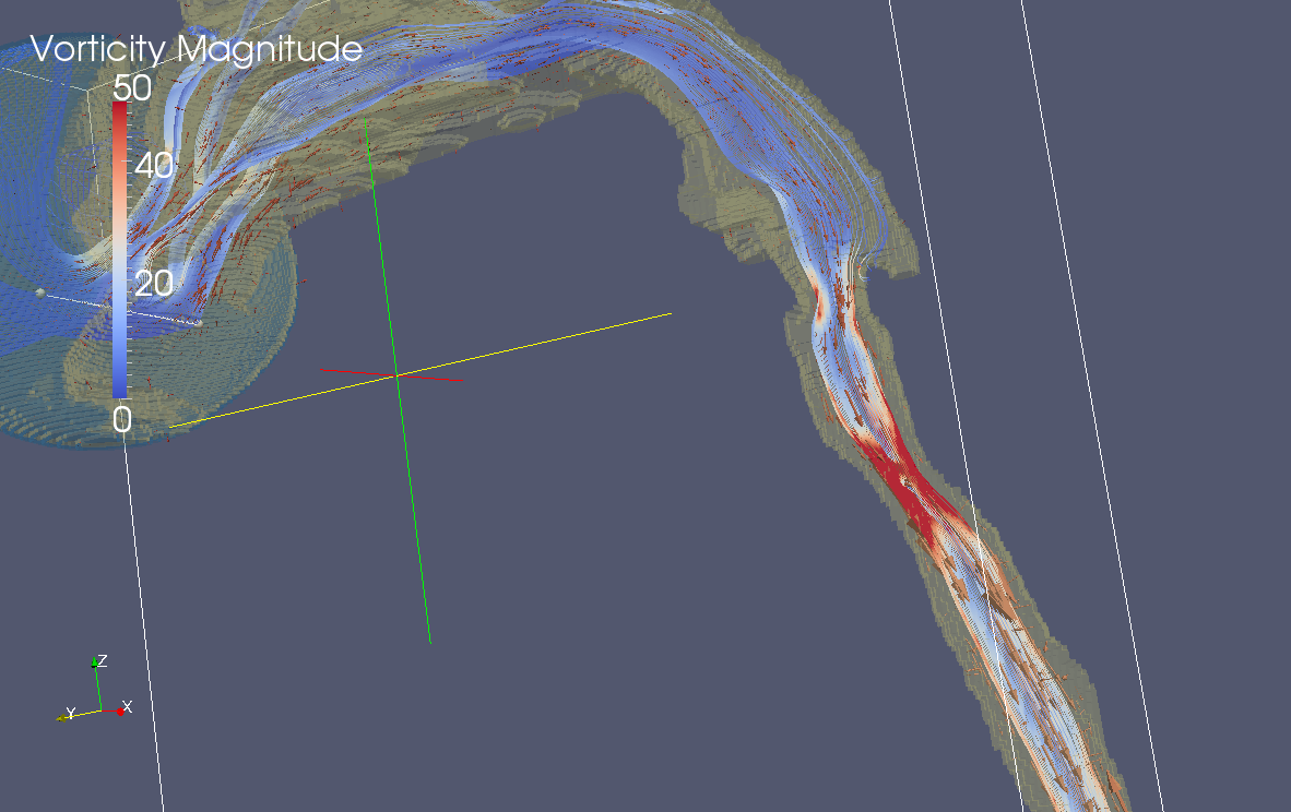
Along with Ron Kikinis, MD, from Brigham and Women’s Hospital and Harvard Medical School, I will present “Evolving themes for HPC in medical imaging.” In particular, we will highlight several successes, missed predictions, and future opportunities for HPC in research and clinical medical imaging, including ongoing work in modeling biological processes spanning molecular interactions, electrical activity modeling, blood flow simulation, and tissue-level anatomic motion assessment. Through our presentation and others, the workshop will identify what we need to link the research and practice of image analysis and visualization in healthcare. I invite you to join the workshop and learn how we can improve the healthcare field. I also invite you to stop by the Kitware booth (#3437) to further discuss how HPC relates to medical image analysis and visualization. At the booth, my team members and I will happily overview both collaboration and employment opportunities.
Beyond medical imaging techniques, Kitware team members will provide insights into the field of HPC and visualization throughout SC16. In particular, they will highlight in situ computation with ParaView Catalyst, which has run on over one million cores. To learn about the variety of activities at which you can find us at the conference, check out our event listing. We look forward to seeing you in Salt Lake City!