
How to Use the SlicerSALT 1.0 Data Importer
The Slicer Shape AnaLysis Toolbox (SlicerSALT) is an open source software framework that helps researchers study shape changes in anatomy. With modules such as Data Importer, the framework enhances the intuitiveness of morphology analysis studies. Data Importer is especially helpful for reviewing the topology of complex anatomical structures. We use it with craniofacial data, as shown below.
You can download synthetic data on Girder such as “case02_allSegments.” The data consists of different label maps with multiple labels, each of which highlight a structure for analysis. Data Importer not only visualizes the cohort of label maps in a dataset, but it visualizes individual labels within each data file. The module also evaluates the shape topology of every label, indicating whether all labels within the cohort have consistent shapes. Since some of SlicerSALT’s methods are constrained to a certain shape topology, Data Importer can help you detect and troubleshoot potential analysis errors in the input data.
To select Data Importer in SlicerSALT, type its name in the magnifying glass in the Modules menu, or browse for it in the dropdown menu.

You can either import data from a directory or from a comma-separated value file. Once the correct input data source has been loaded, use the Import button to load all the input data into the scene.
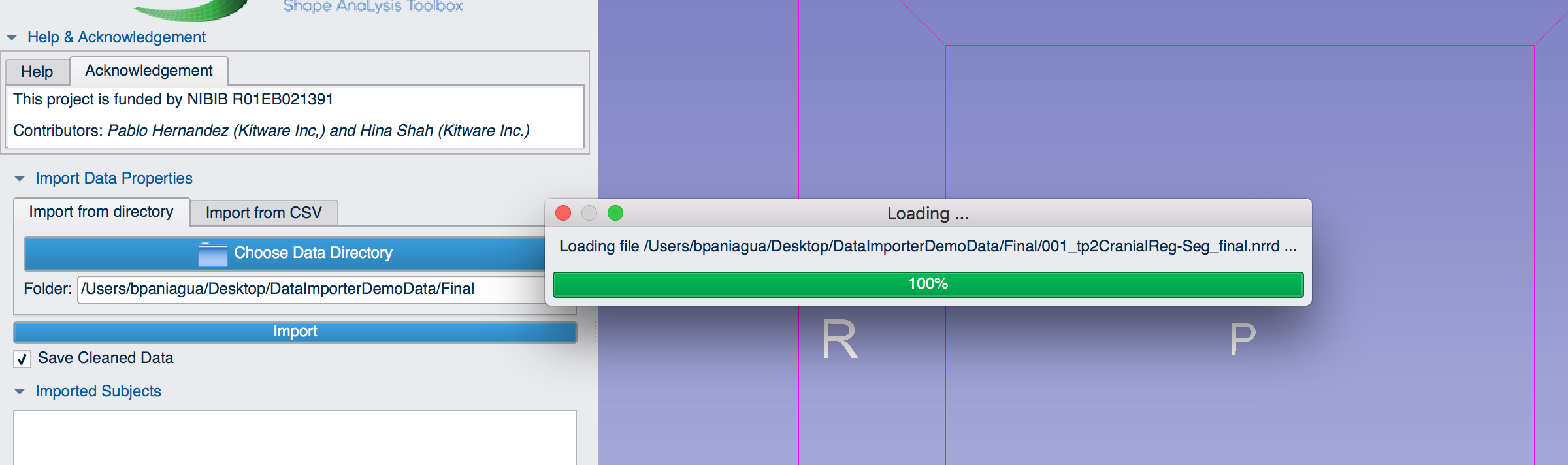
If the data is loading correctly, a dialog will appear.
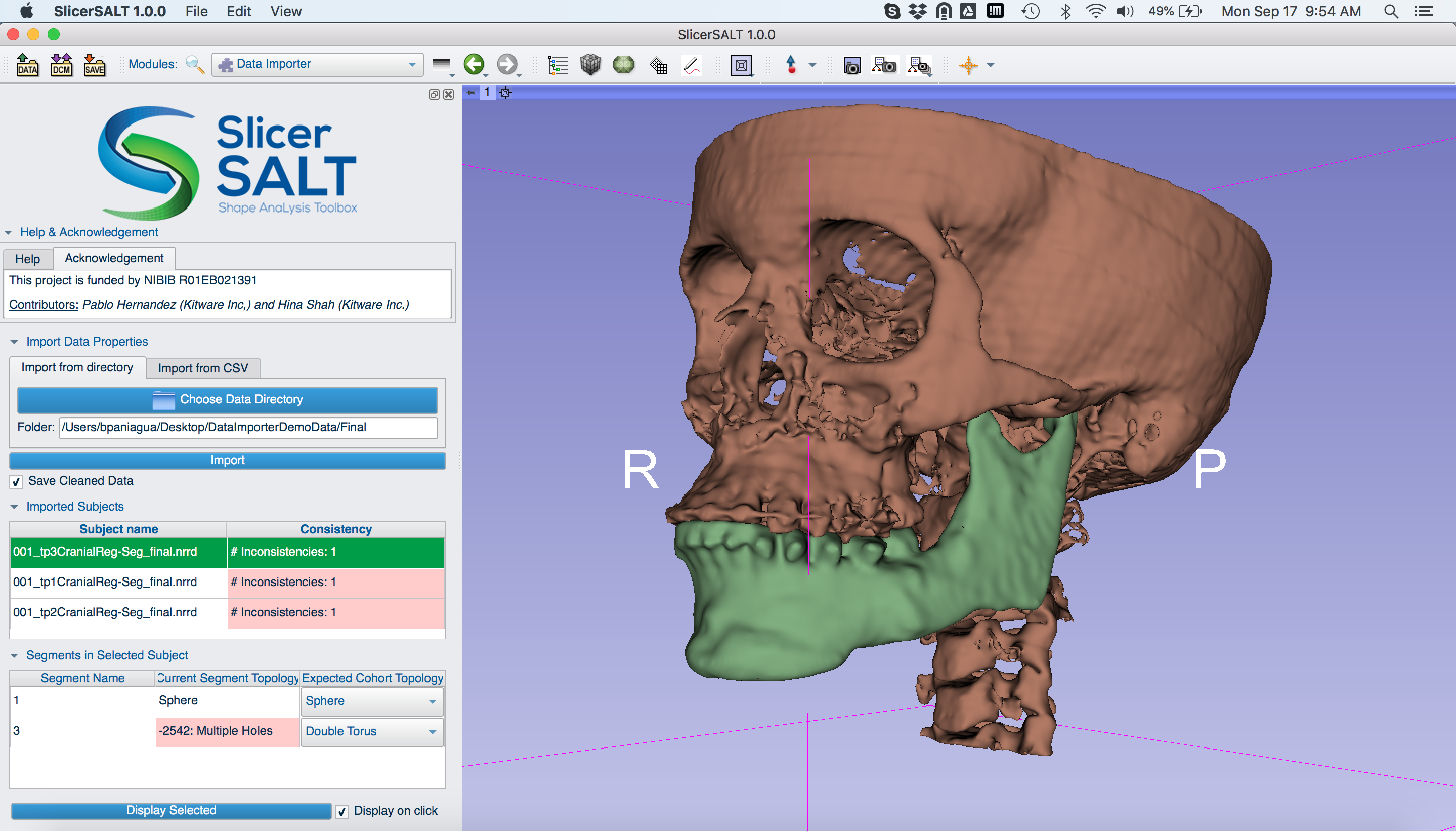
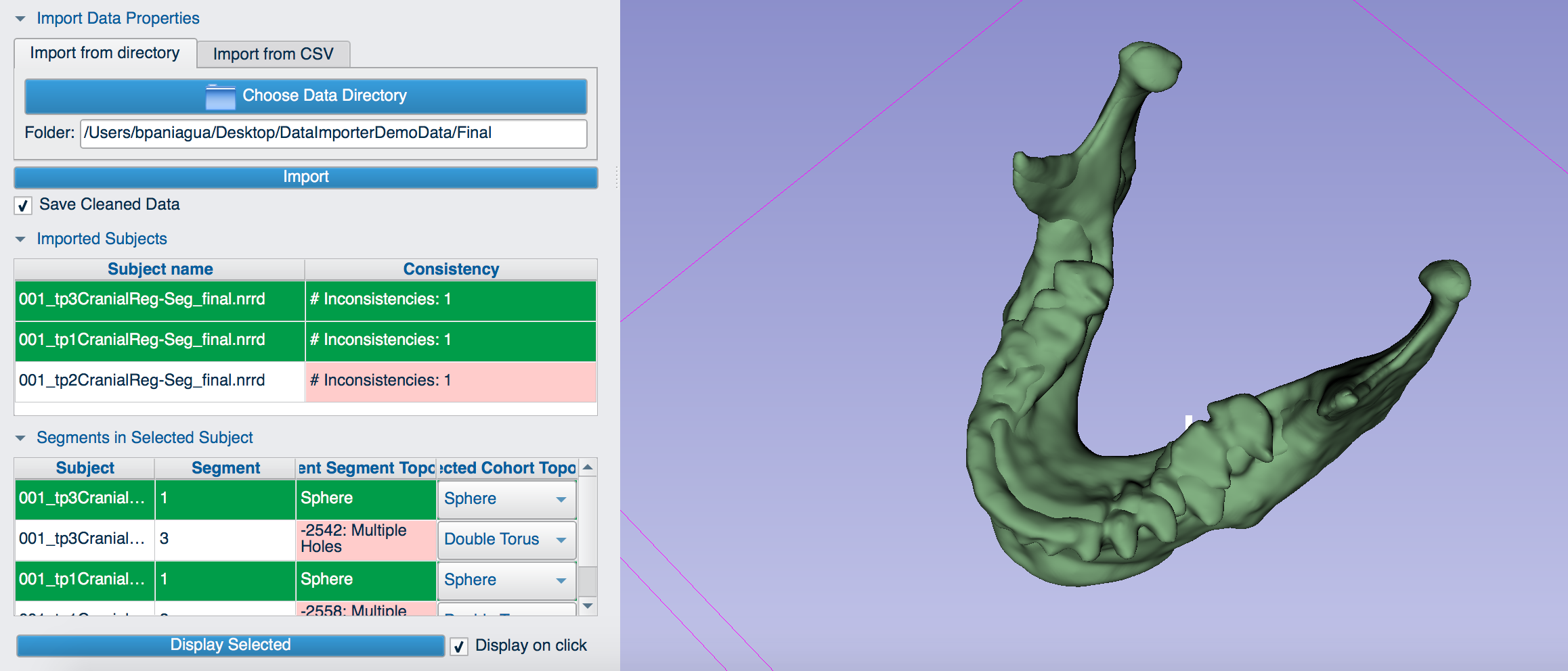
The input data to the Data Importer module is a cohort of segmented anatomies. This means that every case should include the same number of segmented labels. To aid with data quality control, Data Importer has multiple tables: Imported Subjects and Segments in Selected Subject. The module can display each label for a given subject together, depending on the Subjects/Labels selection in the tables.
In our example, we load three time points (subjects) for the same orthognathic surgery patient: one pre-surgical time point and two post-surgical time points. Each time point includes two labels. As the screenshot indicates, the mandible label, is consistent for all subjects. The maxilla label, however, includes inconsistencies. This means that the anatomical structure does not have the same topology in the cohort of different time points.
You can fix the the topology of the structure in a segmentation package. In the future, we plan to extend the Data Importer module to automatically parse the output folders of well-known segmentation programs used in medical image analysis such the FMRIB Software Library, FreeSurfer and Autoseg. None of these platforms currently enable sophisticated shape analysis.
Research reported in this publication was supported by the National Institute Of Biomedical Imaging And Bioengineering of the National Institutes of Health under Award Number R01EB021391. The content is solely the responsibility of the authors and does not necessarily represent the official views of the National Institutes of Health.