
Kitware Receives Funding to Develop an Innovative Method for Quantifying Lung Cancer Markers in Longitudinal CT Studies

Kitware Inc. announces a $202,762 STTR award from the National Institutes of Health to develop a computational method for longitudinal image analysis.
The project is a collaboration between Kitware and three world-class research institutions: Rochester Institute of Technology (RIT), The University of North Carolina at Chapel Hill (UNC), and University of Pittsburgh. It is co-led by Dr. Nathan Cahill of RIT and Dr. Marc Niethammer of UNC Chapel Hill’s Department of Computer Science.
Over this two-year project, Kitware’s research will focus on the continued development of the geometric metamorphosis algorithm that it pioneered with Dr. Niethammer at UNC. This algorithm facilitates longitudinal image analysis by capturing and quantifying pathology-specific changes regarding disease growth and infiltration, while also compensating for background motion in patient scans taken over time. Most current techniques do not distinguish diseases that spread by displacing healthy tissue from diseases that spread by infiltrating healthy tissue; however, distinguishing those types of changes can be vital to disease diagnosis and treatment monitoring. Furthermore, most current techniques do not de-couple background motion (e.g., respiratory motion) from disease change. Therefore, they provide an imprecise estimate of disease change.
While geometric metamorphosis is applicable to the longitudinal study of nearly any type of focal pathology, this project will focus on one of the most challenging and important clinical tasks: the detection and diagnosis of subtle lung lesions that may be pre-cursor to the development of lung cancer. To pursue this clinical goal, the geometric metamorphosis algorithm will be integrated with more accurate models of lung motion developed by Drs. Nathan Cahill and Maria Helguera at RIT. Additionally, Drs. Kyongtae Ty Bae and David Fetzer, Radiology Faculty in the Department of Radiology at the University of Pittsburgh, will provide clinical guidance and data throughout the development and evaluation of these techniques.
“We are excited to be collaborating with such an outstanding team and working to develop a technology that addresses a significant clinical need that has been overlooked by much of the medical imaging research community,” Dr. Stephen Aylward, Kitware’s Senior Director of Operations in North Carolina and project lead for this effort at Kitware, said. “Our successful preliminary work has already inspired others to begin to investigate similar algorithms, and ultimately this body of work will provide clinicians with more sensitive and specific metrics for diagnosing diseases and monitoring treatment efficacy.”
Research reported in this publication was supported by the National Institute Of Biomedical Imaging And Bioengineering of the National Institutes of Health under Award Number R41EB015775. The content is solely the responsibility of the authors and does not necessarily represent the official views of the National Institutes of Health.
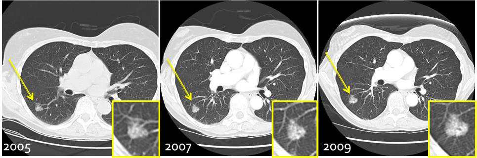
Evolution of a subsolid “ground-glass” lesion over four years.