Kitware Announces the Release of 3D Slicer 4.5 for Medical Image Analysis
Version 4.5 release employs software practices from Kitware to accelerate the progression of research from the lab to the patient’s bedside.
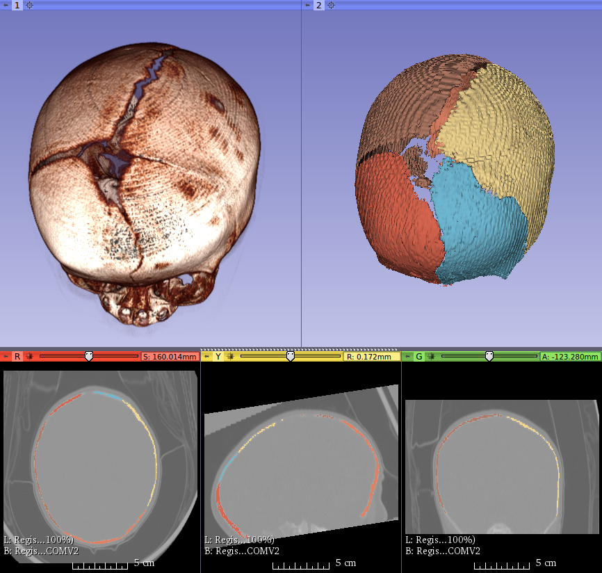
Kitware, Inc., on behalf of the 3D Slicer community, is pleased to announce the release of 3D Slicer version 4.5 in support of basic and applied medical imaging research. 3D Slicer is an industry-leading software application for visualizing and processing medical images from a multitude of medical devices. Researchers have used it to correlate the shape of structures in magnetic resonance imaging (MRI) scans of brains with the risk of schizophrenia, as well as to measure cardiac function from computed tomography (CT).
One of the many strengths of 3D Slicer is its extensible, open-source foundation. The 4.5 release offers over 60 extensions and 150 feature improvements, in addition to performance and stability advancements. Extensions are provided by a community of over 160 researchers from around the world and are free for academic and commercial use. These extensions enable 3D Slicer to import numerous new kinds of medical images and to make precise measurements of a variety of anatomic structures and pathologies. One extension, for example, computes biomarkers for lung disease from CT images.
“Both a full-featured research platform enabling reproducible science and a highly customizable software application meeting rigorous industrial expectations, 3D Slicer is at the forefront of cutting-edge medical research and patient care,” said Jean-Christophe Fillion-Robin, who led the 3D Slicer 4.5 development effort at Kitware.

|
The 4.5 release tightens the integration between 3D Slicer and the Insight Segmentation and Registration Toolkit (ITK). It also takes advantage of recent developments in the Visualization Toolkit (VTK). These toolkits, like 3D Slicer, contain best-in-class scientific methods and employ Kitware’s high-quality software practices, based on CMake, for building and distributing stable applications on Windows, Linux, and MacOS platforms. For a full set of 3D Slicer 4.5’s features, please visit 3D Slicer’s Wiki page.
“3D Slicer is a prime example of the power and integrative potential of ITK, VTK, and CMake,” said Fillion-Robin. “The image and data analysis algorithms and extensions of 3D Slicer are written using ITK, the visualization capabilities of 3D Slicer are provided by VTK, and the cross-platform building and testing of 3D Slicer are managed by CMake. These libraries are industry leaders and are freely available for academic and commercial use within and beyond 3D Slicer.”
To learn how to leverage Kitware’s open-source platforms, including 3D Slicer, ITK, VTK, and CMake, into research, products, and processes, please contact Jean-Christophe Fillion-Robin at kitware(at)kitware.com. To learn more about 3D Slicer, please visit http://www.slicer.org.
3D Slicer is supported, in part, by grants associated with the National Alliance for Medical Image Computing (NA-MIC), the Neuroimage Analysis Center (NAC), the National Center for Image-Guided Therapy (NCIGT), and numerous other National Institutes of Health and government initiatives. Much of the 3D Slicer version 4.5 release was supported by the National Cancer Institute of the National Institutes of Health under Award Number U24CA194354. The content does not necessarily represent the official views of the National Institutes of Health.