Model-to-Image Registration in TubeTK

Registering images (i.e., determine how they align with one another) is a common problem in many domains including medicine, computer vision, and manufacturing. For example, it can be used to monitor changes in a patient over time to assess response to treatment or to align satellite images over time to assess geological changes. Three general approaches to image registration are: (a) image-to-image registration which optimizes the pixel-level match between the images; (b) feature-to-feature image registration in which features are extracted from both images, and then those feature sets are registered; and (c) model-to-image registration in which features (i.e., "models") are extracted from one image and then registration occurs by comuting the fit of the model to the other image. The advantages of model-to-image registration are that it only requires extraction from one image (which saves time and reduces the chance of error), the images can be from different sources with the model acting as a converter, and the model can be used to provide spatially and geometrically focused measures of registration fit which speeds the registration process.
The TubeTK project provides methods for model extraction and model-to-image registration that are applicable to a wide range of data. Furthermore, it contains several specializations for model extraction and model-to-image registration. For example, it has effective methods for modeling tubular structures (e.g., vessels in MRI or CT medical images or roads in satellite images) and for registering models to ultrasound images. For registering models to ultrasound images, TubeTK incorporates knowledge of ultrasound image formation physics into the model-to-image fit measures. Those methods estimate the expected appearance characteristics of the model in the ultrasound data and use that information to tune the spatial and geometric focus of the measures of fit. These specialization enhance the speed and accuracy of the convergence of the registration algorithm.
This work was recently presented by Kitware's Dr. Matt McCormick at the SIIM 2014 conference. For more information, see Matthew M. McCormick, PhD, Kitware; Andinet Enquobahrie, PhD, Kitware; Sharif Razzaque, PhD, InnerOptic; Brian Heaney, InnerOptic; Andrei State, PhD, InnerOptic; Stephen R. Aylward, PhD, Kitware, "USING THE PHYSICS OF ULTRASOUND ACQUISITION IN VESSEL-TO-IMAGE REGISTRATION FOR IMAGE-GUIDED SURGERY" The Society for Imaging Informatics in Medicine annual conference, Long Beach, CA, May 15-17, 2014.
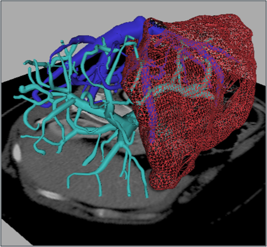
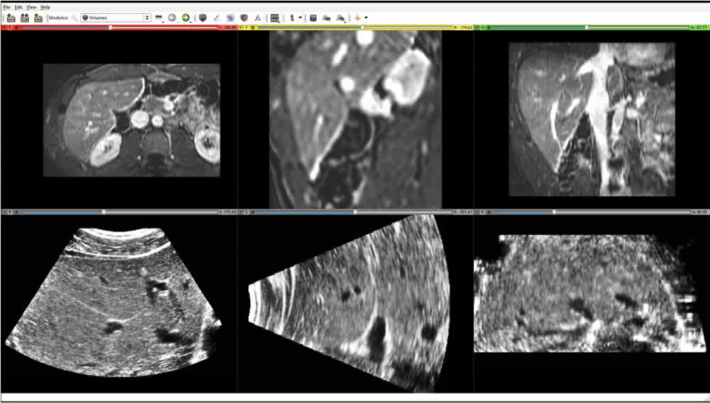
Figure: First image: Models formed using the extraction methods implemented in TubeTK. Second image: Slices through 3D enhanced CT (top row) and 3D ultrasound images (bottom row) that can be registered using the model-to-image registration methods of TubeTK.
To learn more about the TubeTK project and how it improves image registration, watch “Model to Image Registration in TubeTK” and visit http://tubetk.org.